Scientists Identify 'Superspreader' Proteins Linked to Alzheimer's
A new study has used powerful imaging to reveal a subset of Alzheimer's-associated proteins spreading particularly rapidly. These 'superspreaders' may help explain why abnormal clumps of the naturally occurring amyloid beta proteins increase as the debilitating disease progresses.
"This work brings us another step closer to better understanding how these proteins spread in brain tissue of Alzheimer's disease," explains molecular physicist Peter Nirmalraj from the Swiss Federal Laboratories for Materials Science and Technology (EMPA).
Prying the direct cause of Alzheimer's neurological damage from the chaotic molecular tangles in our brains has so far proven extremely difficult. Whether amyloid beta plaques are a cause or a symptom of the disease is also still up for debate.
Laboratory studies have found no evidence these proteins directly damage brain cells, and treatments aimed at these proteins have not proven effective.
Research published this year suggests that while the plaques themselves may not directly cause neuron damage, other molecules that get tangled up with them may be responsible. Regardless, understanding the mechanisms behind the overgrowth of amyloid beta proteins may still lead to better treatments.
In their new study, Nirmalraj and colleagues from the University of Limerick in Ireland used powerful new techniques to watch how amyloid beta proteins join together into long-stranded fibrils that eventually form tangled clumps.

The team observed this activity in a salt solution, which they note is much closer to the natural environment in our brains than other laboratory imaging conditions.
"Conventional methods, such as those based on staining techniques, could alter the morphology and adsorption site of the proteins so that they cannot be analyzed in their natural form," Nirmalraj points out.
Around 250 hours of observation under an atomic force microscope allowed the researchers to spy something unusual.
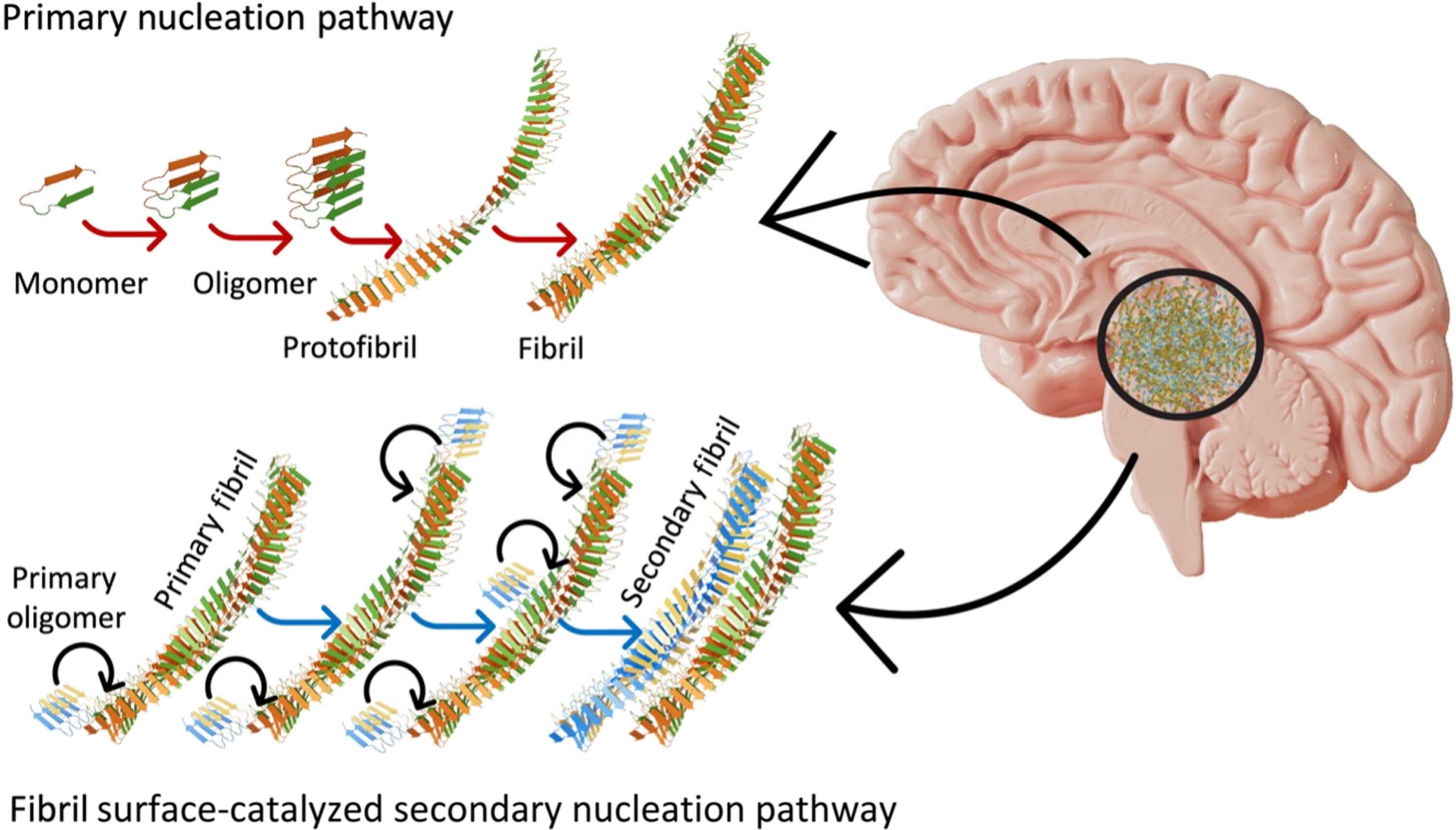
A specific flavor of amyloid beta proteins fold in such a way that their edges are extra reactive, as seen in the bright areas of the above image. This means they more readily accumulate additional building blocks of themselves – extending their string-like fibrils through the brain quicker than other types of amyloid beta do.
"A small population of fibrils that displayed higher surface catalytic activity was identified as superspreaders," the team writes in their paper.
These superspreaders, amyloid beta 42, form only as secondary structures after an initial amyloid beta fibril has been created (see the diagram below).

Nirmalraj and team clarify the shape and size of these protein clumps, but details of their structure are yet to be resolved. These details include if and how amyloid beta 42 differs chemically from other amyloid beta proteins, and what chemistry drives the growth of these secondary structures.
Previous research has found that as amyloid beta proteins aggregate in the brain, people suffer from increasingly distressing symptoms such as memory loss, impulsive behavior, anxiety, and confusion. This is still just an association and the direct mechanism for brain damage in dementia is not yet understood.
A vast complexity of changes occur over decades in the brains and bodies of people developing neurodegeneration. So there are other theories that require continued exploration too, such as Alzheimer's as an autoimmune disease.
This research was published in Science Advances.


